Welcome to Prager-Khoutorsky Lab

About Us

         We are interested in understanding integrative mechanisms by which the brain regulates basic functions of the body, such as hunger, thirst, and hormonal levels. The area of the brain involved in this control is the hypothalamus, which coordinates the neuroendocrine system, cardiovascular system, energy metabolism, fluid homeostasis, and sleep. We are particularly interested in mechanisms by which specific hypothalamic areas communicate with the periphery to detect the levels of circulating molecules (metabolites, ions, and hormones) and to generate adaptive responses to adjust the physiological needs of the organism. Disruption of these mechanisms can lead to pathological conditions such as hypertension, obesity, and diabetes.

         Our lab is located in downtown Montreal, one of the most unique, cosmopolitan, and vibrant cities in North America. The city boasts exceptional cultural diversity and has been ranked as the best city in the world for students in 2016.       

  We are currently looking for motivated and talented candidates for graduate and undergraduate research as well as postdoc positions.

Research Interests

1. Cellular and molecular mechanisms underlying plasma sodium detection by the brain and their role in the salt-sensitive hypertension. Specifically, we study a function of specialized osmosensory neurons that are activated by increased levels of sodium in the blood. These neurons release antidiuretic hormone vasopressin to stabilize the levels of sodium and water in the circulation. We investigate the role of unique cytoskeletal structures and signalling molecules featured by these neurons, and study how these elements are affected by high dietary salt, mediating hyperactivation of osmosensory neurons, and thereby contributing to hypertension.

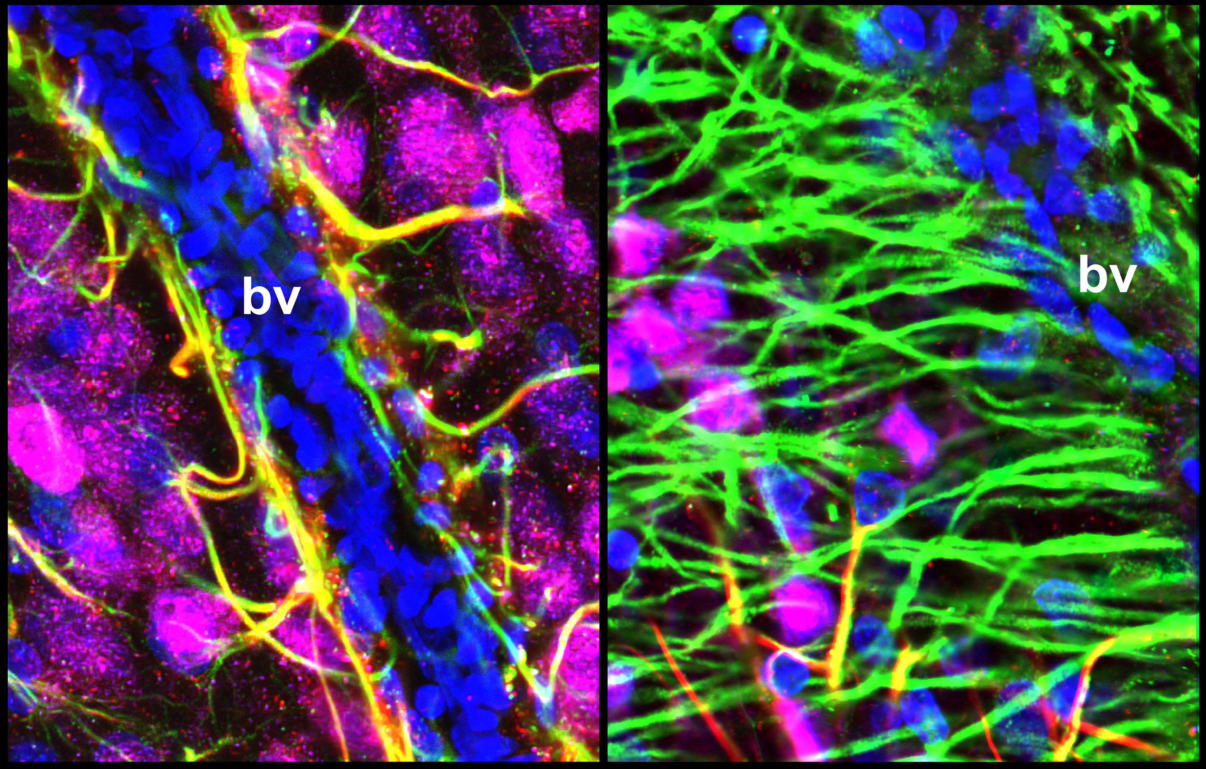
2. Cellular and molecular mechanisms underlying the communication between the brain and the peripheral circulation: the role of non-neuronal cells (tanycytes, astrocytes, pericytes, and endothelial cells) in the regulation of the blood brain barrier. Metabolites, hormones and other circulating molecules cannot freely penetrate into the brain due to the existence of the blood brain barrier, which isolates the brain from molecules found in the peripheral circulation. Specialized small brain regions in the hypothalamus (called circumventricular organs) are lacking the complete blood brain barrier. Therefore, blood-borne circulating molecules can partially access these areas to influence the activity of local neurons, which in turn generate neuronal responses by mediating hormonal release, promoting behaviors (e.g. by triggering thirst or hunger), or activating autonomic nerve system to modulate cardiovascular system and renal function. Our goal is to understand the cellular and molecular mechanisms that regulate the access of peripheral signals to neurons in the circumventricular organs and to study functions of non-neuronal cells (tanycytes, astrocytes, pericytes, and endothelial cells) in this dialogue between the periphery and the central nervous system.

3. Cellular and molecular mechanisms underlying the regulation of the blood brain barrier in health and metabolic disorders. Tanycytes have been proposed to serve as gatekeepers mediating the communication between the brain and the body at hypothalamic circumventricular organs. Tanycytes can adjust by altering the functional and structural organization of the blood-hypothalamus barrier in response to nutritional status of an individual (e.g. fasting vs feeding). We investigate molecular and cellular mechanisms that underlie these plastic changes, and study how disruption of these processes leads to metabolic disorders promoting obesity, diabetes, and hypertension.

To address these questions, we use a variety of techniques:

  • Patch clamp electrophysiological recordings (brain slices, dissociated cells)
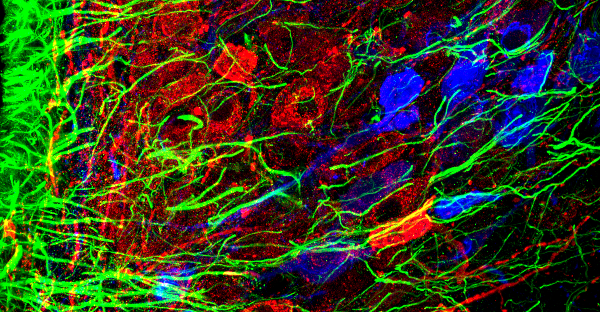
  • Super-resolution imaging
  • Live-cell imaging (calcium, cytoskeleton, and signaling molecules)
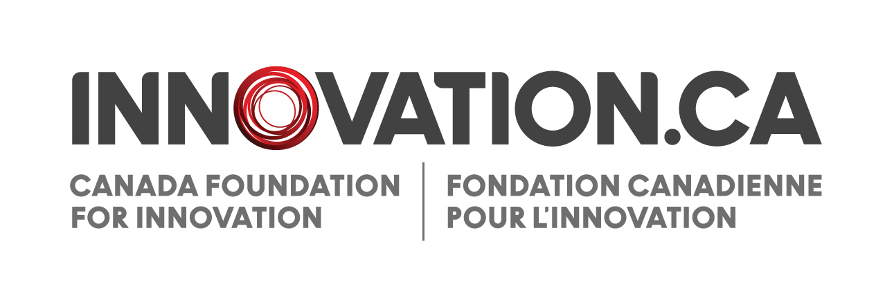
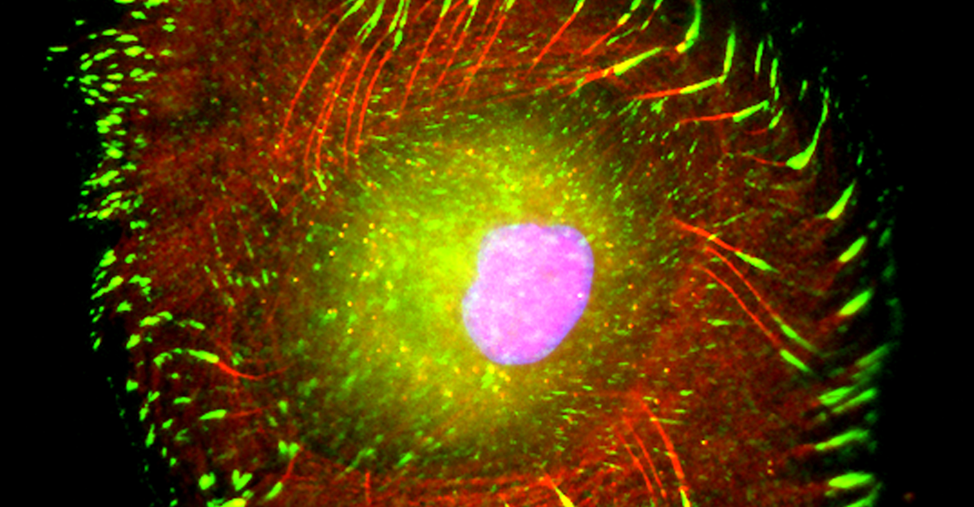
  • Immunohistochemistry, histology, and neuronal-glial-vasculature morphometry
  • Hemodynamic measurements (blood pressure and heart rate)
  • Animal models of human diseases

Current Group Members

Masha

Masha Prager-Khoutorsky

Principal Investigator
Assistant Professor
Jieyi

Jieyi Yang


Lab Manager


Ning Gu


Research Associate

Project: Molecular analysis of sodium detection

Olena

Olena Makashova


Postdoctoral fellow

Project: Hypothalamic microglia & osmoregulation

Pierre-Marie Chevillard


Postdoctoral Fellow

Project: Hypothalamic adult neurogenesis

Amirah

Amirah-Iman
Hicks

Grad Student

Project: Regulation of the blood brain barrier

Simona Kobrinsky


Grad Student

Project: Vasopressin in salt-dependent hypertension

Susana

Susana
Puche Saud

Grad Student

Project: Glia-neuron interactions & hypothalamus

Chris

Chris Chen


Grad Student

Project:

Celeste

Celeste Laporte


Grad Student

Project:

Yining

Yining Zhou


Undergrad Student
Brenna

Brenna Shimizu


Undergrad Student

Alumni :
David Levi, Betty Rodriguez, Melissande Bry, Saijel Patel, Jeremy Baylou, Jasper Sim, Mishaal Hukamdad, Suleima Jacob-Tomas, Zsuzsanna Barad, Graham Lean, Anna Zarzycki, Alberto Sobrero, Misha Hubacek, Safiya Rizwan, Suijian Zhou, Vanessa Josey, Banruo Li.

Selected Publications

  • Hicks AI, Prager-Khoutorsky M. Neuronal culprits of sickness behaviours. Nature. 2022 Sep 609(7928):679-680. doi: 10.1038/d41586-022-02321-7
  • Tansley S, Gu N, Guzmán AU, Cai W, Wong C, Lister K, Muñoz-Pino E, Yousefpour N, Roome RB, Heal J, Wu N, Castonguay A, Lean G, Muir EM, Kania A, Prager-Khoutorsky M, Zhang J, Gkogkas CG, Fawcett JW, Diatchenko L, Ribeiro-da-Silva A, De Koninck Y, Mogil JS, Khoutorsky A. Microglia-mediated degradation of perineuronal nets promotes pain. Science. 2022 May 26. doi: 10.1126/science.abl6773.
  • Wong C, Barkai O, Wang F, Thörn Pérez C, Lev S, Cai W, Tansley S, Yousefpour N, Hooshmandi M, Lister KC, Latif M, Cuello AC, Prager-Khoutorsky M , Mogil JS, Séguéla P, De Koninck Y, Ribeiro-da-Silva A, Binshtok AM, Khoutorsky A. mTORC2 mediates structural plasticity in distal nociceptive endings that contributes to pain hypersensitivity following inflammation. J Clin Invest. 2022 May 17:e152635. doi: 10.1172/JCI152635.
  • Tansley S, Uttam S, Ureña Guzmán A, Yaqubi M, Pacis A, Parisien M, Deamond H, Wong C, Rabau O, Brown N, Haglund L, Ouellet J, Santaguida C, Ribeiro-da-Silva A, Tahmasebi S, Prager-Khoutorsky M, Ragoussis J, Zhang J, Salter MW, Diatchenko L, Healy LM, Mogil JS, Khoutorsky A. Single-cell RNA sequencing reveals time- and sex-specific responses of mouse spinal cord microglia to peripheral nerve injury and links ApoE to chronic pain. Nat Commun. 2022 Feb 11;13(1):843. doi: 10.1038/s41467-022-28473-8.
  • Rodríguez-Cortés B, Hurtado-Alvarado G, Martínez-Gómez Luis R, León-Mercado LA, Prager-Khoutorsky M, Buijs RM. Suprachiasmatic nucleus-mediated glucose entry into the arcuate nucleus determines the daily rhythm in blood glycemia. Current Biology. February 28, 2022. doi:10.1016/j.cub.2021.12.039
  • Prager-Khoutorsky M. (2021) Cytoskeletal Organization and Plasticity in Magnocellular Neurons. In: Grinevich V., Dobolyi Á. (eds) Neuroanatomy of Neuroendocrine Systems. Masterclass in Neuroendocrinology, vol 12. Springer, Cham. https://doi.org/10.1007/978-3-030-86630-3_5
  • Hicks AI, Kobrinsky S, Zhou S, Yang J, Prager-Khoutorsky M. Anatomical Organization of the Rat Subfornical Organ. Front Cell Neurosci. 2021 Sep 6;15:691711. doi: 10.3389/fncel.2021.691711.
  • Levi, DI., Wyrosdic, JC., Hicks, AI., Andrade , MA., Toney, GM., Prager-Khoutorsky, M*, Bourque, CW*. High dietary salt amplifies osmoresponsiveness in vasopressin-releasing neurons. (* co-last authors). Cell Rep. 2021 Mar 16;34(11):108866. doi: 10.1016/j.celrep.2021.108866.
  • Barad, Z., Jacob-Tomas, S., Sobrero, A., Lean, G., Hicks, AI., Yang, J., Choe KY., Prager-Khoutorsky, M. Unique Organization of actin cytoskeleton in magnocellular vasopressin neurons in normal conditions and in response to salt-loading. eNeuro. 2020 March 24 ENEURO.0351-19.2020. doi:10.1523/ENEURO.0351-19.2020.
  • Hicks, AI., Barad, Z, Sobrero, A., Lean, A., Jacob-Thomas, S., Yang, J., Choe, KY., Prager-Khoutorsky, M. Effects of Salt Loading on the Organization of Microtubules in Rat Magnocellular Vasopressin Neurons. J Neuroendocrinol. Epub: 2019 Nov 28. doi:10.1111/jne.12817
  • Ciura, S., Prager-Khoutorsky, M., Thirouin, ZS., Wyrosdic, JC., Olson, JE., Liedtke, W., and Bourque, CW. (2018). TRPV4 mediates hypotonic inhibition of central osmosensory neurons via taurine gliotransmission. Cell Rep. 2018 May 22;23(8):2245-2253
  • Uttam, S., Wong, C., Amorim, IS., Jafarnej, SM., Tansley, SN., Yang, J., Prager-Khoutorsky, M., Mogil, JS., Gkogkas, CG., Khoutorsky, A. Translational profiling of dorsal root ganglia and spinal cord in a mouse model of neuropathic pain. Neurobiology of Pain. Epub: 2018 Apr 18. doi: 10.1016/j.ynpai.2018.04.001.
  • Prager-Khoutorsky, M. (2017) Mechanosensing in hypothalamic osmosensory neurons. Semin Cell Dev Biol. doi: 10.1016/j.semcdb.2017.06.006
  • Prager-Khoutorsky, M., Choe, KY., Levi, DI., Bourque CW. (2017) Role of Vasopressin in rat models of salt-dependent hypertension.Curr Hypertens Rep, 19(5):42.
  • Khoutorsky, A*., Sorge, R*., Prager-Khoutorsky, M*., Gkogkas, C., Martin, L., Pitcher, M., Austin, JS., Pawlowski, SA., Longo, G., Sharif-Naeini, R., Ribeiro-da-Silva, A., Bourque, CW., Cervero, F., Mogil, J and Sonenberg, N. (2016) Cellular stress response pathway controls thermal nociception via translational regulation of TRPV1. (* co-first authors). PNAS 113(42):11949-11954.
  • Zaelzer, C., Hua, P., Prager-Khoutorsky, M., Ciura, S., Voisin, DL., Liedtke, W., and Bourque, CW. (2015) ΔN−TRPV1 encodes a molecular integrator of physiological temperature and hypertonic stress. Cell Rep,13(1):23-30.
  • Prager-Khoutorsky, M. and Bourque, C.W. (2015) Anatomical organization of the rat organum vasculosum lamina terminalis. Am J Physiol Regul Integr Comp Physiol, 309(4): 324-37.
  • Prager-Khoutorsky, M. and Bourque, C.W. (2015) Mechanical basis of osmosensory transduction in magnocellular neurosecretory neurons of the rat supraoptic nucleus. J Neuroendocrinol, 27(6):507-15.
  • Prager-Khoutorsky M., Khoutorsky A, and Bourque CW. (2014) Unique interweaved microtubule scaffold mediates osmosensory transduction via physical interaction with TRPV1. Neuron, 83(4):866-78.
  • Prager-Khoutorsky M., Lichtenshtein A, Krishnan R., Rajendran K., Mayo A., Kam Z., Geiger B. and Bershadsky AD. (2011). Fibroblast polarization is a matrix rigidity-dependent process controlled by focal adhesion mechanosensing. Nat Cell Biol, Nov 13;13(12):1457-65.
  • Prager-Khoutorsky M. and Bourque CW. (2010). Osmosensation in vasopressin neurons: changing actin density to optimize function. Trends Neurosci. Feb:33(2):76-83.

For a complete list of publications, visit PubMed.

Biosketch

I received my PhD from the Hebrew University of Jerusalem, Israel, where I investigated the interplay between cytoskeleton and plasma membrane dynamics (endo- and exocytosis) under normal conditions and during axonal regeneration. I characterized the important role of crosstalk between membrane recycling and cytoskeleton dynamics in regulation of neuronal morphology.

For my first postdoctoral training, I joined one of the world’s leading groups studying cytoskeletal mechanisms of mechanotransduction in non-neuronal cells supervised by Drs. Benjamin Geiger and Alexander Bershadsky at The Weizmann Institute of Science, Israel. To identify new signaling molecules regulating mechanosensation, I designed a new microscopy-based screening assay capable of revealing proteins involved in mechanotransduction and discovered 20 tyrosine kinases involved in cellular sensing of the mechanical environment. This work (Prager-Khoutorsky et al, Nature Cell Biology, 2011) provided new insights into fundamental processes such as cell polarization and motility.

For the second postdoctoralship, I joined the laboratory of Dr. Charles Bourque at McGill University, to study the cellular mechanisms underlying another form of mechanosensation - osmosensation. In the Bourque lab, I explored the roles of mechanosensitive channels, cytoskeletal elements, and the interplay between them in mediating mechanotransduction in osmosensory neurons. Additionally, I developed a new methodology that enables to visualize subcellular cytoskeletal networks in neurons in-situ using super-resolution microscopy. I discovered a unique intertwined scaffold of microtubules present exclusively in osmosensory neurons. These microtubules physically interact with the transduction channel at the surface of the osmosensory neurons and push-activates the channel mediating activation of the neurons. These findings (Prager-Khoutorsky et al, Neuron, 2014) provided the first evidence supporting a role for microtubules in mechanotransduction and expanded the understanding of mechanisms by which the brain monitors and corrects the body’s hydration state.

In addition, I became interested in the physiology of another hypothalamic osmosensory nucleus called OVLT, which is a unique brain area that lacks a blood brain barrier, and thus can sense molecules circulating in the blood. The OVLT is involved in regulation of vital functions of the organism, including body sodium homeostasis, cardiovascular and neuroendocrine systems, sexual and reproductive behaviors, thermoregulation, and immune responses. Detailed analysis of this region resulted in discovery of a previously undescribed large population of specialized ependymal cells, called tanycytes, which create a dense network of tanycytic processes, embedding local neurons and are possibly involved in the regulation of their activity. Understanding the function of the OVLT in mediating the bi-directional communication between the brain and the body and exploring the role of tanycytes in physiological and pathological conditions are integral parts of my long-term research program.

Have Any Questions? Contact Us

Department of Physiology,
McGill University

McIntyre Medical Building, Rm 1230 
3655 Prom. Sir-William-Osler 
Montreal, QC, Canada, H3G 1Y6 
Canada 
Lab Tel: 514-398-4565

Masha Prager-Khoutorsky 
McIntyre Medical Building, Room 1237 
Office Tel: 514-398-1818
masha.prager-khoutorsky@mcgill.ca